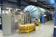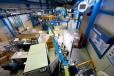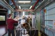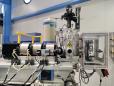
High-energy heavy ion microprobe
The high-energy heavy-ion microprobe is used for the characterisation or modification of material properties at depths from approximately 1 micrometre to maximum depths of up to 500 micrometres from the material surface.