Welcome to MEX!
We're now taking user proposals for beam time on MEX1 and MEX2.
To find out more, including our current capabilities, visit our User Wiki. To learn more about submitting a proposal for beamtime, visit the facility access page.
We're now taking user proposals for beam time on MEX1 and MEX2.
To find out more, including our current capabilities, visit our User Wiki. To learn more about submitting a proposal for beamtime, visit the facility access page.
October 2023
The microprobe has landed!
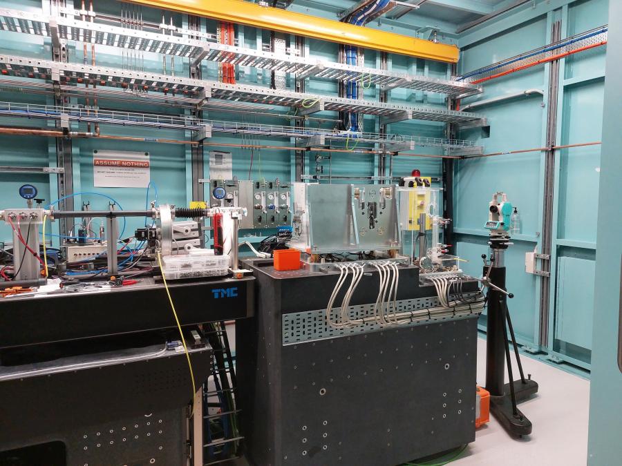
The September shutdown was a whirlwind of activity at MEX1, with the installation of the microprobe! After many months of building and testing, the microprobe was finally ready to move into its forever home at the end of the MEX1 endstation in Hutch B.
Once the microprobe was installed, Beamline Scientist Dr Simon James made quick work of aligning the components and achieving not only first light on the new equipment, but collecting the first spectra off an iron foil!
This achievement was a wonderful collaborative effort between the Science, the Mechanical Engineering and Surveying, Controls Engineering, Electrical Technicians, and Project Management teams. Well done all!
Commissioning work will continue over the coming months to optimise the microprobe to the conditions required by the user community.

April 2023
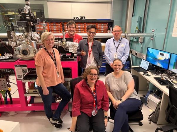
On April 26, Associate Professor Rosalie Hocking, Dr Ngoc Khang Dinh, and PhD candidates Brittany Kerr and Jaydon Meilak were the first merit users on the MEX-2 beamline! The team from Swinburne University of Technology used the beam time to investigate the sulfur chemistry of catalysts for the hydrogen evolution reaction. They were supported by Beamline Scientists Dr Bruce Cowie and Dr Valerie Mitchell.
This milestone comes after achievement of first light on MEX-2 in December 2022, and is a credit to the hard work and dedication of the entire MEX team. Work will continue over the coming months and years to roll out MEX’s full capability.
Now that both MEX-1 and MEX-2 are operational beamlines, capability updates will be posted on our User Wiki.
December 2022
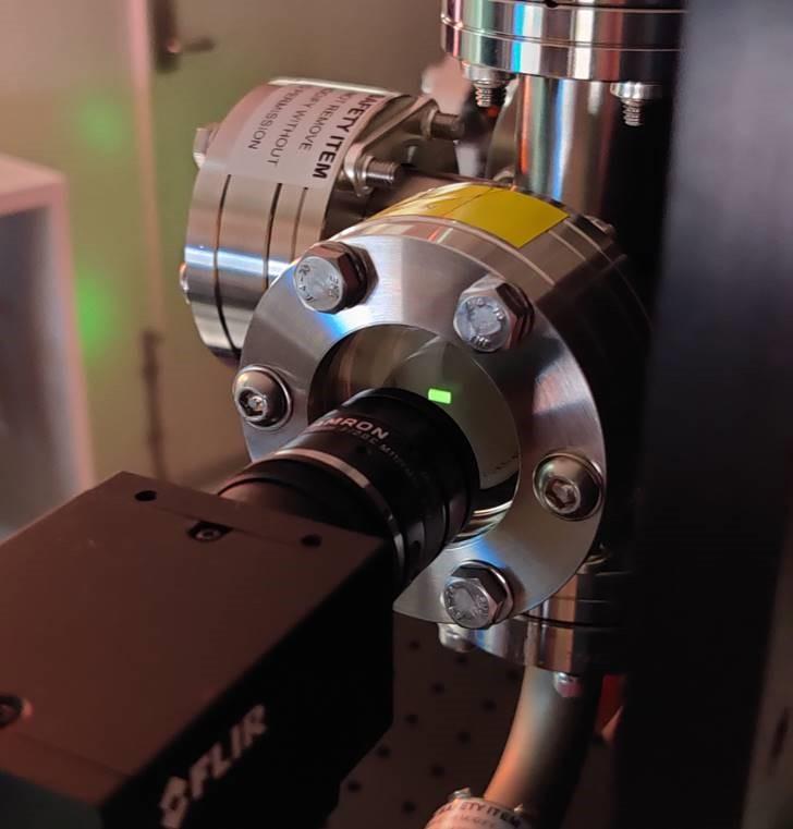
The MEX team were very excited to achieve first light - where we thread synchrotron light through our beamline for the first time - on the MEX2 beamline on December 7!
The MEX2 endstation doesn't need to be enclosed in a hutch, so we were able to look directly at the beam on a phosphor screen through a lead glass window - see the green rectangle in the image below.
The team will take a much-needed break over the holidays and return in 2023 to continue commissioning in preparation for first users on MEX2 in April.
November 2022
We've had yet another huge milestone this month at MEX, and this one is the biggest yet!
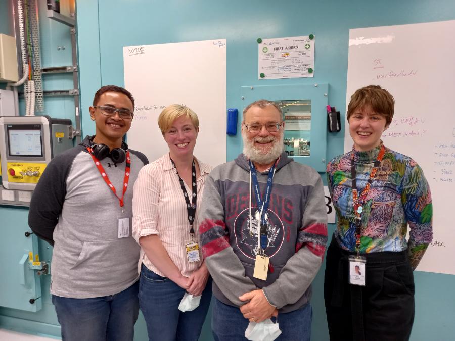
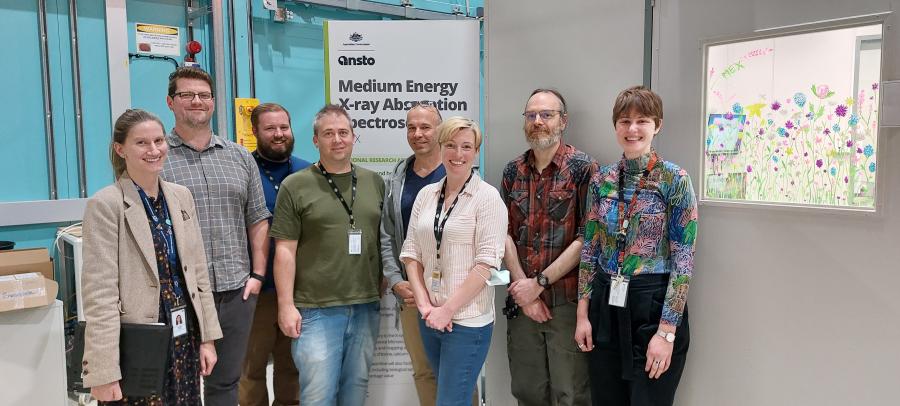
On 23 November, Professor Brendan Kennedy and PhD candidate Ahmadi Permana were the first merit users on the MEX-1 beamline. The team from The University of Sydney used the beam time to investigate the local structure and oxidation state of perovskite materials and were very pleased with the data they collected. This milestone comes after achievement of first light in June 2022, and is a credit to the hard work and dedication of the entire MEX team. Work will continue over the coming months and years to roll out MEX’s full capability.
October 2022
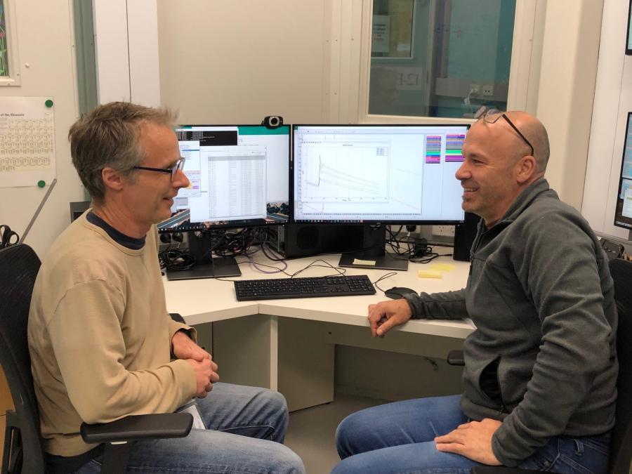
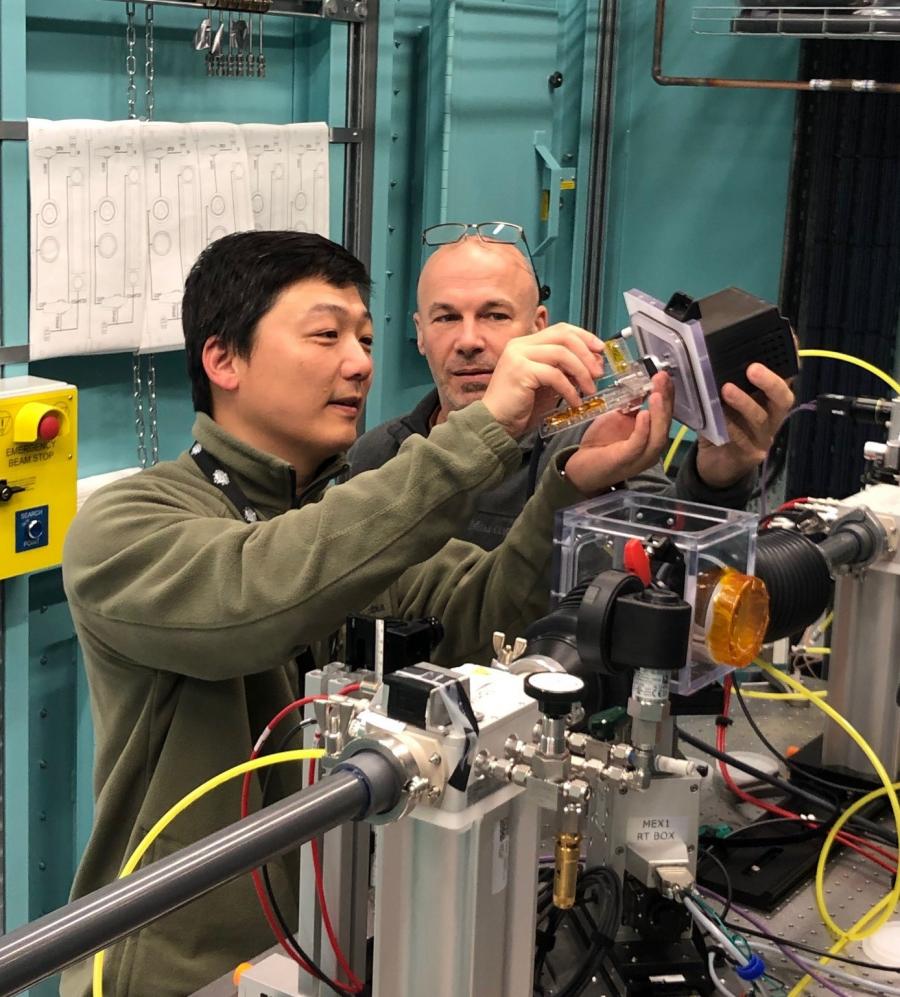
Huge news, MEX fans! This month we had our very first expert users on MEX1! Dr Bernt Johannessen, Senior Scientist at the Australian Synchrotron’s X-ray Absorption Spectroscopy (XAS) beamline, was our first expert user in early October. Bernt was followed by Principal Scientist from our Powder Diffraction (PD) beamline, Dr Qinfen Gu, who ran an experiment on MEX1 a week later.
Both scientists collected excellent data and provided feedback on their experience which is invaluable for the MEX team to prepare for merit users in late November.
In other exciting news, this month we’re delighted to welcome Dr Valerie Mitchell to the MEX science team! Valerie’s background is in organic chemistry, and she’s joining MEX on a secondment from the XAS Beamline.
September 2022
Throughout September, the MEX team has been working to characterise and understand the MEX1 beamline optics, including:
All these components are important for the reliable operation of the beamline, as our goal is to ensure that users are able to consistently measure quality x-ray absorption spectra from their samples.
This is tedious, detailed, and careful work to prepare for trial experiments with expert users throughout October.
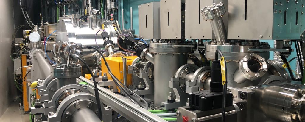
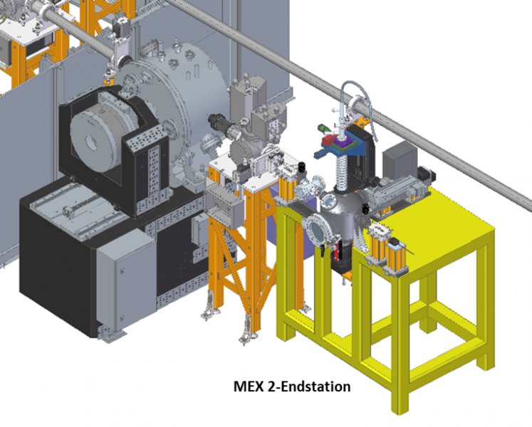
While this flurry of activity has been happening at MEX1, the engineering and technical teams have been hard at work on the final assembly of the MEX2 endstation (image below) ahead of first light in November.

August 2022
In the first two weeks of August, scientists tested and characterised the ion chambers for MEX1 and the microprobe. Ion chambers are gas-based x-ray detectors. The beam interacts with the gas in the ion chamber to form ions and electrons, which are then detected as an electric current. We’ll place our ion chambers on our MEX1 endstation before the sample, after the sample, and after the reference foil to measure the incident beam. The MEX science team will continue characterising the ion chambers after the shutdown, which started on 22 August and ends on 12 September.
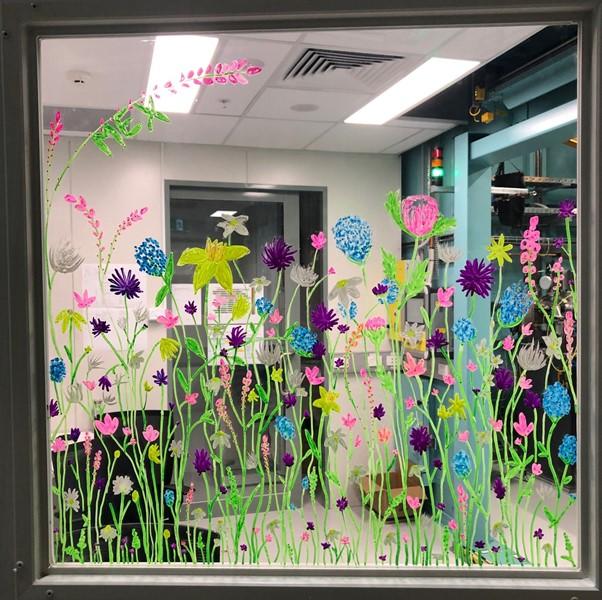
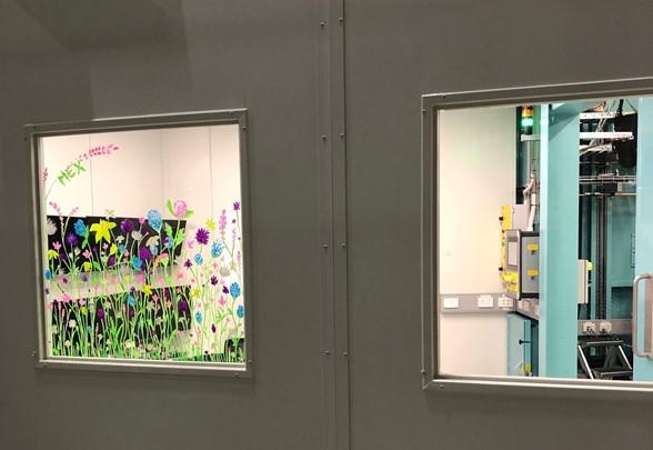
Spring has sprung in Melbourne and at the MEX1 Beamline! With the arrival of shutdown, engineers and technicians have taken over the show to do maintenance on the accelerator and beamline. Meanwhile, the scientists have time to work on their… more creative pursuits (see below).
July 2022
It’s been over a month since we achieved first light at the MEX1 beamline, and we’re still on a first light high! Last month we shared footage of this huge milestone from the user cabin, but in the video below you’ll see what first light looked like from inside the beamline.
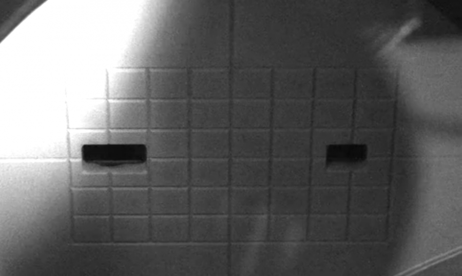
Since first light, we’ve been calibrating and tuning beamline components, and learning how to run the beamline using the computers in our user cabin. We call this process – where we're calibrating equipment with beam in the beamline but are not yet ready for users – hot commissioning (as opposed to cold commissioning, where we were calibrating equipment without beam).
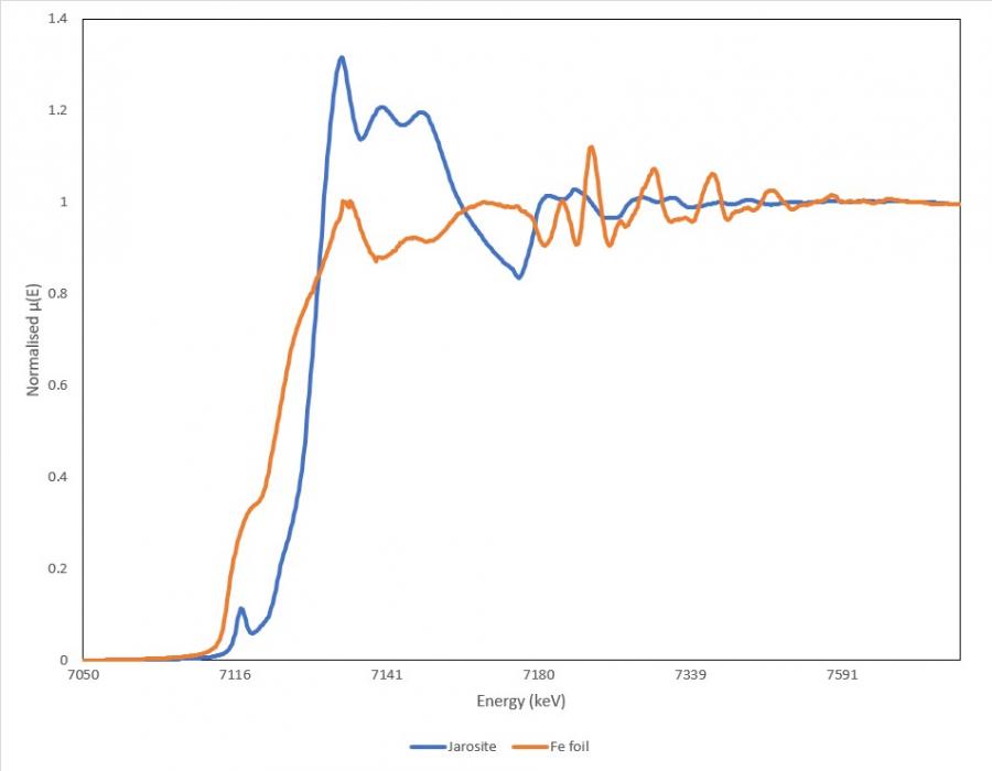
Earlier in the month, we had an opportunity to take MEX1 for a spin on some samples containing iron oxide minerals, which were provided by a Synchrotron user. We’re pretty happy with the results, especially considering the beamline isn’t fully configured yet. Check out the spectra from this experiment in the graph below.
Through the hot commissioning process this month, we’ve investigated key components of the photon delivery system. The photon delivery system is the equipment that focuses, tunes, and shapes the beam before it arrives at users’ samples (at the end station). Next month our focus will shift to characterising the end station, including setting up our brand-new fast ion chambers.
June 2022 - First light!
After months of preparing documents, testing equipment, and getting approvals signed off, the MEX1 Beamline achieved first light on Sunday June 12! First light is when we open the shutter to the synchrotron’s storage ring and allow synchrotron light to enter the beamline for the first time. Folks, it’s kind of a big deal.
Like all good dramas, in the days leading up to June 12, there was a hitch! The synchrotron’s electron gun needed to have a part replaced over that weekend. Although the Accelerator Team could replace this part while keeping current in the storage ring for us to use (if you have no idea what any of these parts are or how a synchrotron works – check out this simple and entertaining explainer by MEX Beamline Scientist Dr Emily Finch), this meant that if for some reason we tripped a safety switch and cut off the beam (which happens all the time!!) while trying to get first light, we wouldn’t be able to get the beam back until the repairs were complete. Basically, we had one shot at first light and if we messed it up we wouldn’t be able to try again until the next week.
So on the morning of Sunday June 12, the MEX team and friends were all gathered in the MEX1 user cabin, ready to open the shutter. There was tense but excited silence as Dr Chris Glover, MEX Lead Scientist, gathered everyone around and pressed the button to open the shutter. We all flicked our heads from the button to the computer monitor, where cameras were trained on the front end of the beamline to watch for the beam of light coming in.
Suddenly, the camera screen lit up bright white – it was over saturated! As we turned down the gains on the camera, a glorious scene came into view: two rectangles of light shining on the front-end mask (the entrance to the beamlines) – MEX first light! It was no NASA Control Room celebration, but it was a great moment and there were certainly little yelps of ‘hooray!’ and ‘yes!’ and ‘there it is!’.
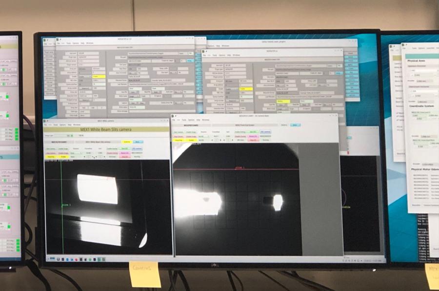
‘What’s next?’ you may ask. Throughout that day the MEX scientists threaded the beam of light through the many components of the MEX1 beamline, all the way to the experimental end station – that’s 30 metres worth of equipment! This is a very complex task, but it was made easier by the fantastic work the engineers and technicians had done in the lead up to first light to ensure the equipment was in the perfect position to make the scientists’ job as easy as possible.
Over the coming weeks and months, the MEX team will work on tuning and calibrating the beamline components to ensure we’re getting the best data possible once we’re ready to take users.
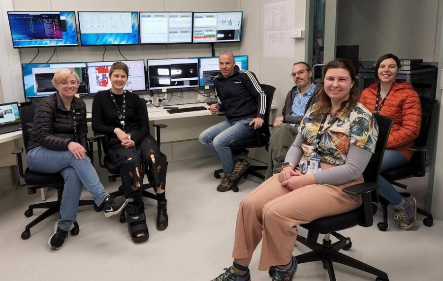
MEX and XAS staff with first light on the monitor in the background.
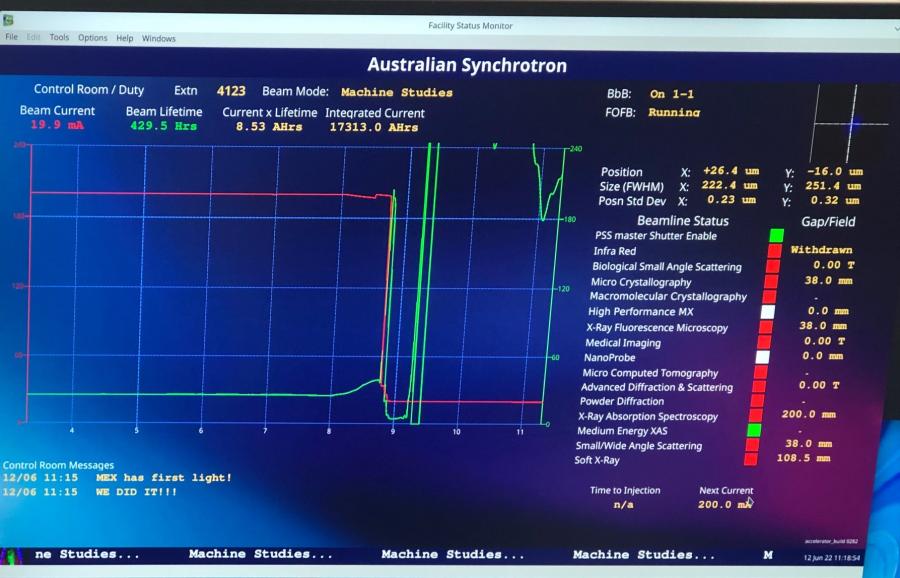
The Facility Status Monitor showing the MEX front end shutter open for the first time, and celebratory messages from the Control Room.
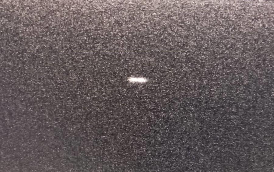
First glimpse of the beam in the MEX-1 Experimental hutch (the far end of the beamline).
May 2022
Welcome back, MEX fans!
We’ve now moved the MEX1 table into its home in the hutch, and started putting together our MEX1 endstation. The endstation is the equipment MEX1 users will use to mount their samples and conduct experiments.
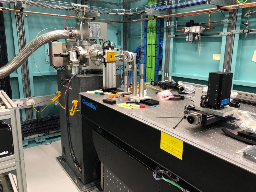
The MEX1 endstation table has been moved into the hutch and is starting to be populated with equipment for the MEX1 endstation.
We’ve also been working with our amazing Scientific Computing and Controls Engineering teams to finalise our graphical user interfaces – the way we (and you!) will operate the beamlines from our user cabins.
We’re now getting close to first light – where we open the shutter to the synchrotron’s stored beam and let synchrotron light into the beamline. This is a huge step in the commissioning process, and will be happening next month! In the meantime, we’ll be testing and calibrating our equipment to make sure it’s ready for this milestone.
March 2022
Okay MEX fans, somehow it’s a quarter of the way through the year now...? I guess that happened while we were busy doing COOL MEX THINGS!
We have now installed both double crystal monochromators (DCMs; remember these? No? Scroll down to the June 2021 update!) into their forever homes in the MEX hutch (MEX1 DCM) and user cabin (MEX2 DCM). We don’t like to have favourites here at MEX, but the DCMs are arguably the most important components of the beamline, so it’s very exciting for us to have these in place.
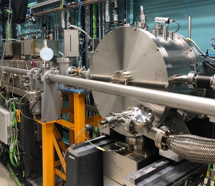
The MEX1 DCM installed in Hutch A. The DCM looks like a large silver cylinder lying on its side behind the MEX2 beam pipe.
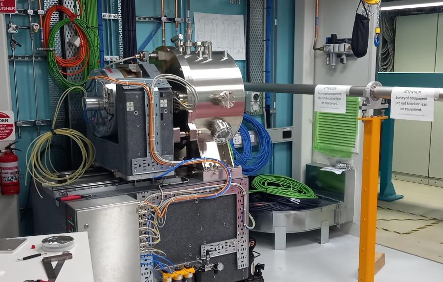
The MEX2 DCM installed in the MEX2 user cabin. The DCMs sit on big blocks of synthetic rock.
We’re also completely delighted that the micro-focusing system for MEX's microprobe passed site acceptance testing. The MEX microprobe will use a small beam (roughly 12 times smaller than the width of a human hair) to study chemical speciation at the micro-scale. Now we’ll move onto developing the rest of the microprobe, starting with the sample positioning system.
And saving the best until last: The 18th International Conference on X-Ray Absorption and Fine Structure (XAFS2022) will be happening online and in person in Sydney in July. The best part of this, in our entirely biased opinion, is that the Australian Synchrotron will be running a satellite workshop as part of XAFS2022, featuring presentations from MEX scientists and a tour of the brand new MEX beamline! Holy smokes!! Expressions of interest are now open at https://xafs2022.org/. We can’t wait to see you down at the beamline!
February 2022
A belated happy new year, MEXers!
After a well-deserved break over the holiday period, the MEX team are back on site and have had a busy start to the year.
The user cabins are finished and ready for us to move into – check them out in the images below. Future users, welcome to your home while doing experiments at MEX!
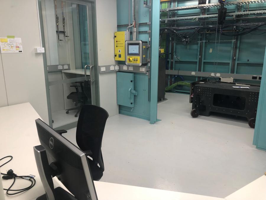
The MEX1 user cabin looking over the user desks towards the sample preparation room and the MEX1 hutch (hutch B).
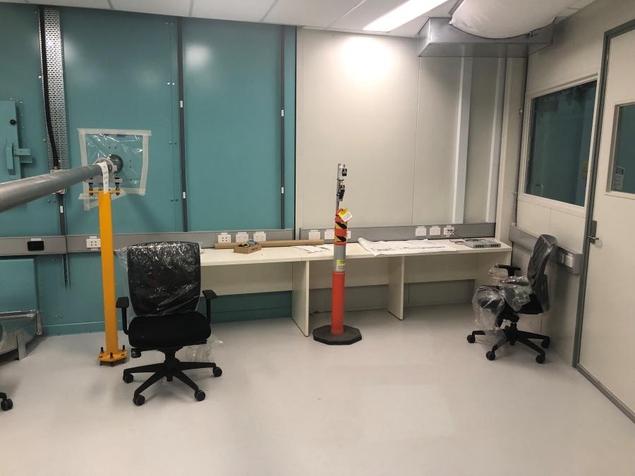
The MEX2 user cabin looking towards the user computer desks (downstream).
Our focusing mirror for MEX1 arrived in January, ready for our team to install in early February. The mirror arrived in a long grey box which protected it during transport. The team took the mirror out of this box in the clean room upon arrival to check that the mirror wasn’t damaged during transport.
Photos of the MEX1 focusing mirror in the Australian Synchrotron clean room upon its arrival inside its box for transport (top) and with the box removed (bottom).
The mirror surface is visible from the bottom, so MEX Lead Scientist, Dr Chris Glover, slid under the mirror to check the mirror surface was clean and unscratched.
Dr Chris Glover and Senior Mechanical Technician Jason Worthensohn inspecting the mirror surface.
This is what he saw.
The surface of the mirror, as seen from below.
The mirror was clean, unscratched, and ready to be installed!
We then moved the mirror into the hutch, carefully removed all the transport locks, installed it, and set up the motion controls for all the motors that move the mirror.
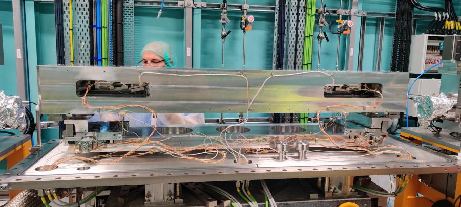
The mirror is in place in the hutch, and Dr Chris Glover is pictured in the process of removing transport locks.
All went well with the installation – a great success!
November 2021
Welcome back, MEXers!
November has brought a fantastic atmosphere back to the Australian Synchrotron - summer is on our doorstep, our faces have emerged from behind masks (mostly), Synchrotron staff and users have returned to site, and commissioning is ramping up at the MEX beamline.
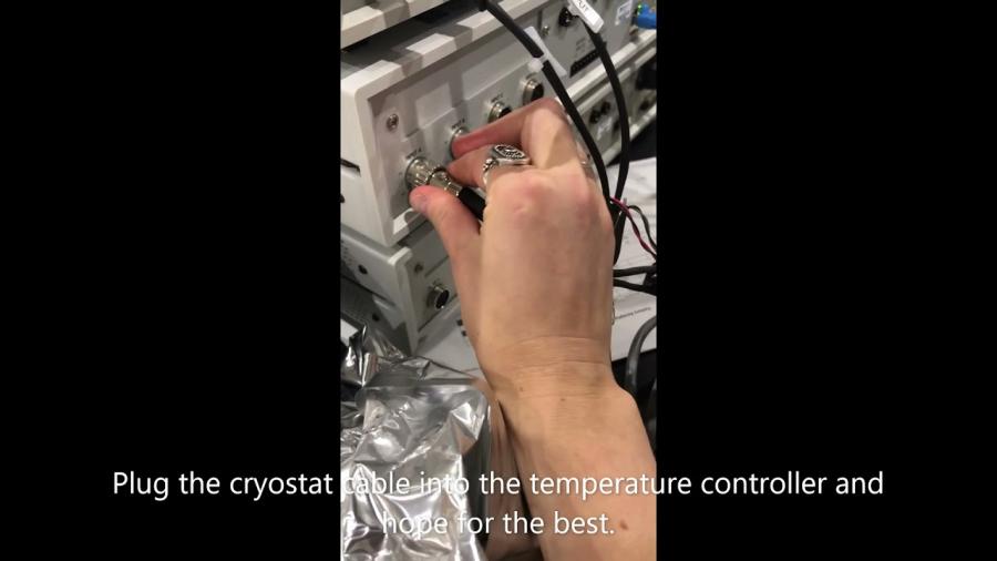
The MEX team have been busy testing out all the equipment that has arrived over the last few months. When this goes well, it involves plugging in one bit of equipment into another and seeing something good happen - see the short video below for a great example of this.
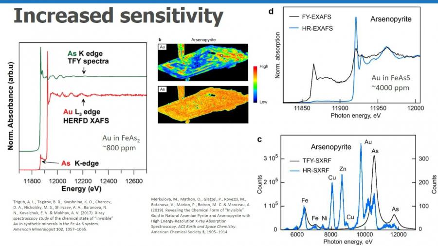
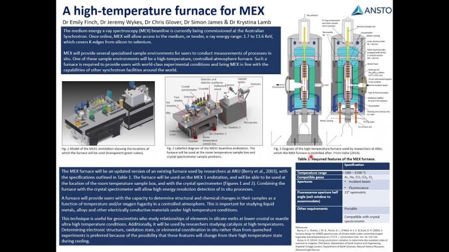
The 2021 ANSTO User Meeting was held on November 24-26. MEX Scientists presented on the crystal spectrometer and the specialised samples environments that MEX will provide for users. The poster presentations for these are below.
October 2021
Melbourne is finally out of lockdown forever (we hope) - After 3 months in our sixth covid lockdown, Melbourne is finally emerging like a plumped-up-on-homemade-sourdough and ungroomed but glorious butterfly emerging from its cocoon.
Now that lockdown is over, the MEX team can start shifting back to working on-site, which means we can dive deeper into commissioning our beamline equipment. November will be busy and exciting!
In the meantime, you can learn more about the MEX beamline and our progress from the recorded talk added in the Technical Information and Specifications section below from Dr Chris Glover, MEX Lead Scientist. Chris recorded this talk for the XAFS2021 conference earlier this year. Enjoy!
September 2021
September has been a quiet month on site. Melbourne is in lockdown due to a COVID outbreak, construction sites are temporarily shut down, and permits are required to carry out essential work on site.
The MEX team has been using this time to test equipment, order parts, get through paperwork, and prepare for the coming months post-lockdown when we'll be starting commissioning without beam.
August 2021
August saw the completion of the MEX photon delivery system installation – the main components of the beamline. MEX engineers and scientists will now spend a few weeks tuning and programming beamline parts to prepare for commissioning in the coming months.

Looking upstream (towards the storage ring) along the PDS in Hutch A.
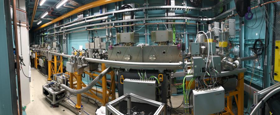
MEX PDS view from the door of Hutch A. Upstream (towards the storage ring) is to the left of the image, and downstream (towards the end stations) is to the right.
In late August, the MEX scientists presented at a joint two-day workshop with the X-ray Absorption Spectroscopy (XAS) beamline team. On the first day of the workshop, MEX scientists discussed the beamline rationale, commissioning timeline, technical setup, and equipment capabilities. We also heard from the XAS team and Synchrotron users about XAS experiments and the exciting science that will be enabled by MEX. The slides from this workshop are available at the event website here.
July 2021
What a difference a month makes! July began with empty MEX hutches, and is ending with Hutch A packed to the rafters. Three engineers from Axilon – a German company that builds accelerator and X-ray instrumentation – have been busy installing MEX’s photon delivery system (PDS).
The PDS is the first part of the beamline the X-rays see when they come out from the storage ring tunnel and into the hutches. It tunes the size, shape, focus, and energy of the X-ray beam. The main components of the PDS include mirrors, double crystal monochromators, and slits.
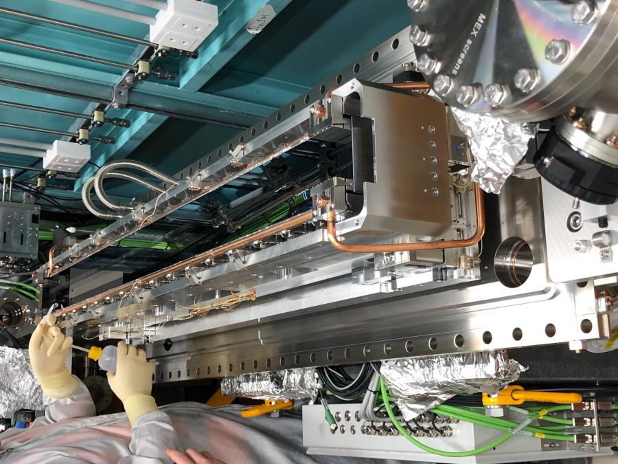
Engineers preparing one of the MEX1 mirrors to be enclosed and put under vacuum.
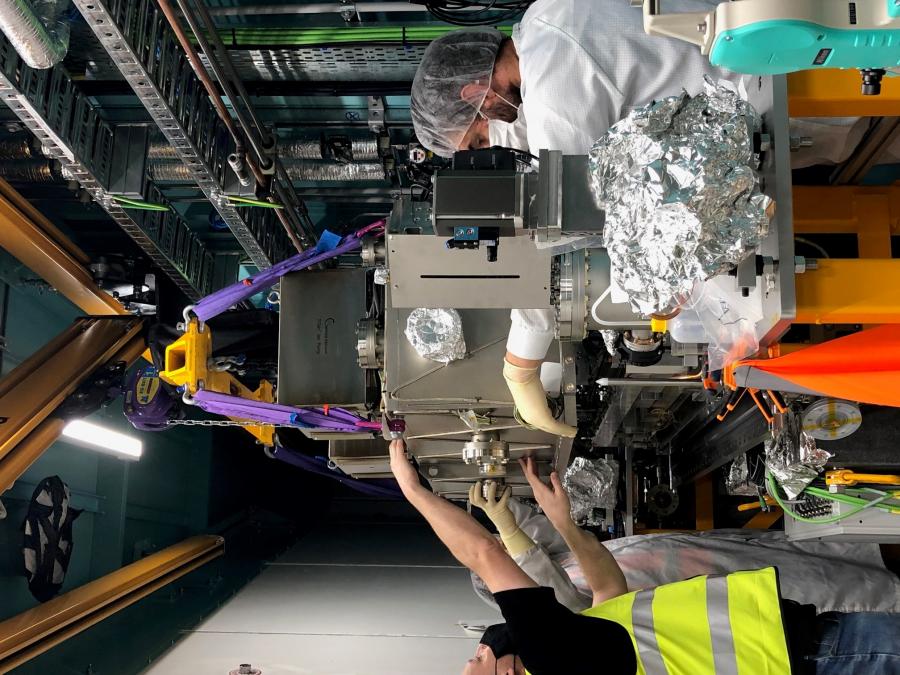
Lowering the MEX1 mirror cover over the mirror using the hutch crane.
MEX scientists tested out the crystal spectrometer on the XAS Beamline earlier this month (for a refresher on the crystal spectrometer, check out the March and May 2021 updates). The goal was to obtain good spectral data from a sample containing mercury, testing out each of the crystals in the five crystal mounts on the spectrometer.
The experiment was a great success! You can see the first high energy resolution fluorescence detection (HERFD) spectra (red) obtained from the crystal spectrometer compared to conventional X-ray absorption near edge structure (XANES) spectra (blue) in the image below.
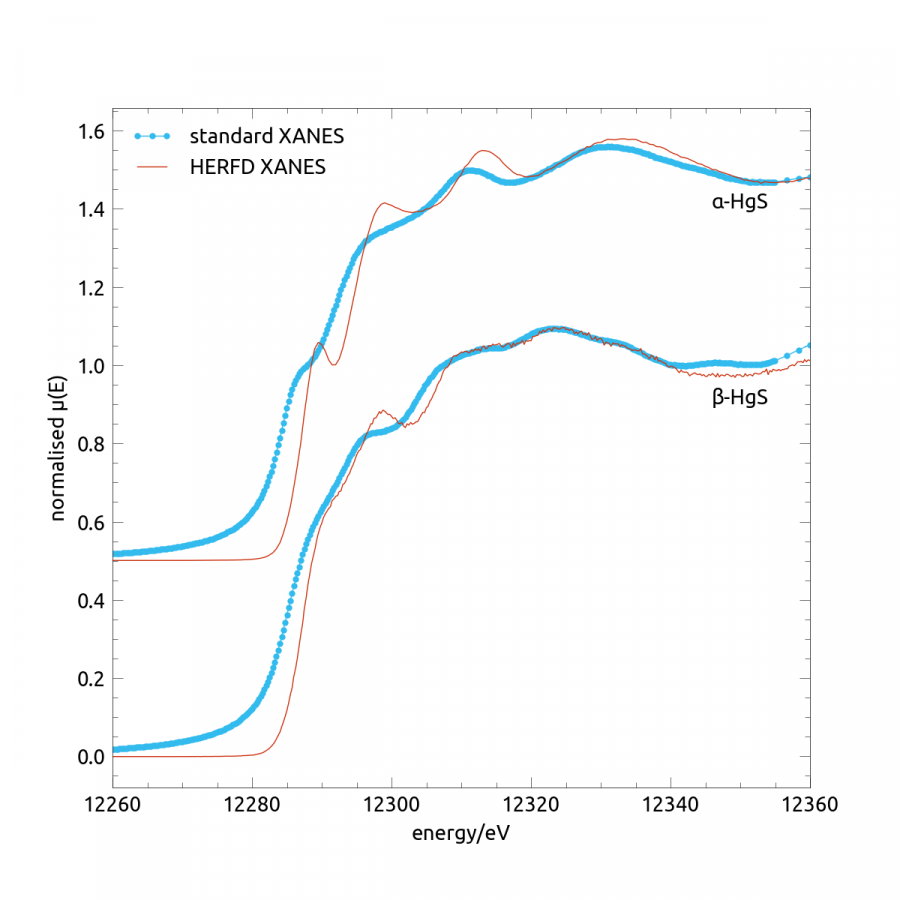
Graph comparing spectra obtained using the crystal spectrometer (HERFD; red) and from conventional XANES (blue).
June 2021
In early June, Rick Ford, an engineer from Instrument Design Technology Ltd, arrived to assemble MEX’s double crystal monochromators (DCMs).
Rick travels around the world assembling and installing synchrotron beamline components (job goals!), including part of the XAS beamline here at the Australian Synchrotron.
The DCMs consist of two sets of two crystals (Si(111) for MEX1; Si(111) and InSb(111) for MEX 2). The crystals are aligned so that the beam hits the first crystal set, is diffracted to the second crystal set, and from there is diffracted to continue along the beamline. The angle of the crystals relative to the beam defines the energy of the beam, so the DCM is one of the most important components of the beamline.
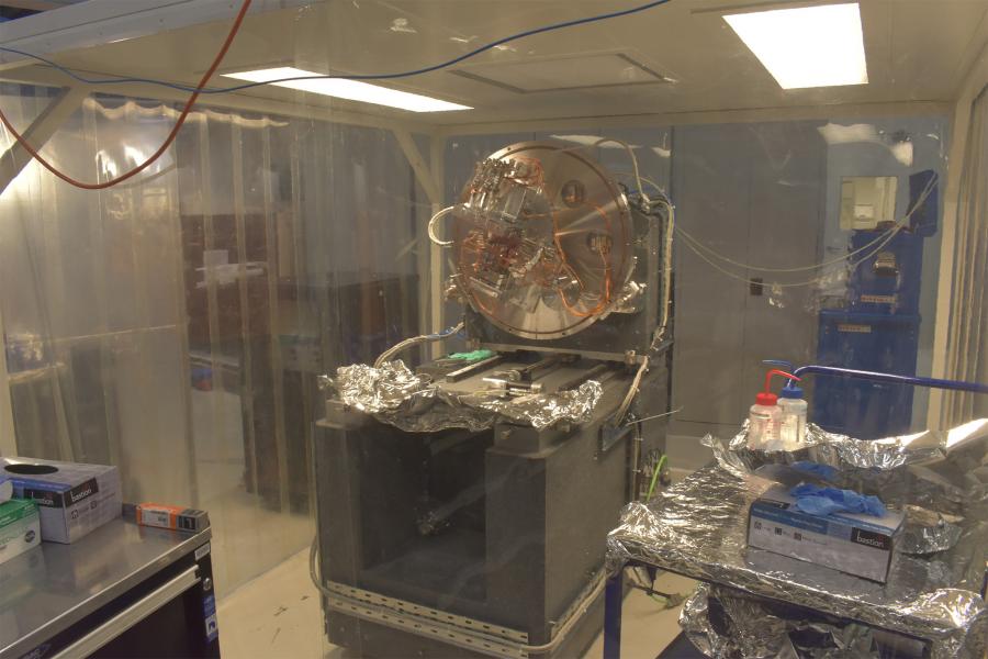
The DCM in a pop-up clean room for assembly.
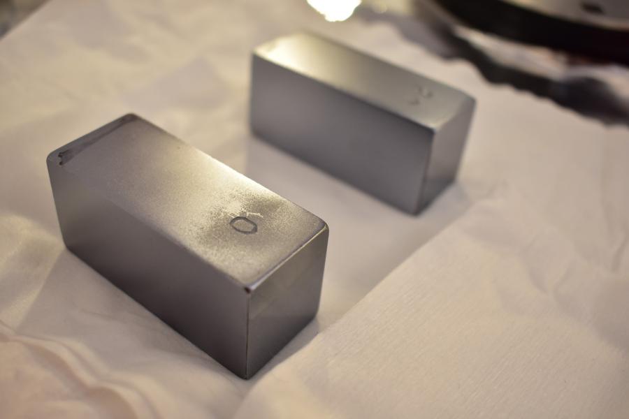
MEX DCM Si(111) crystals.
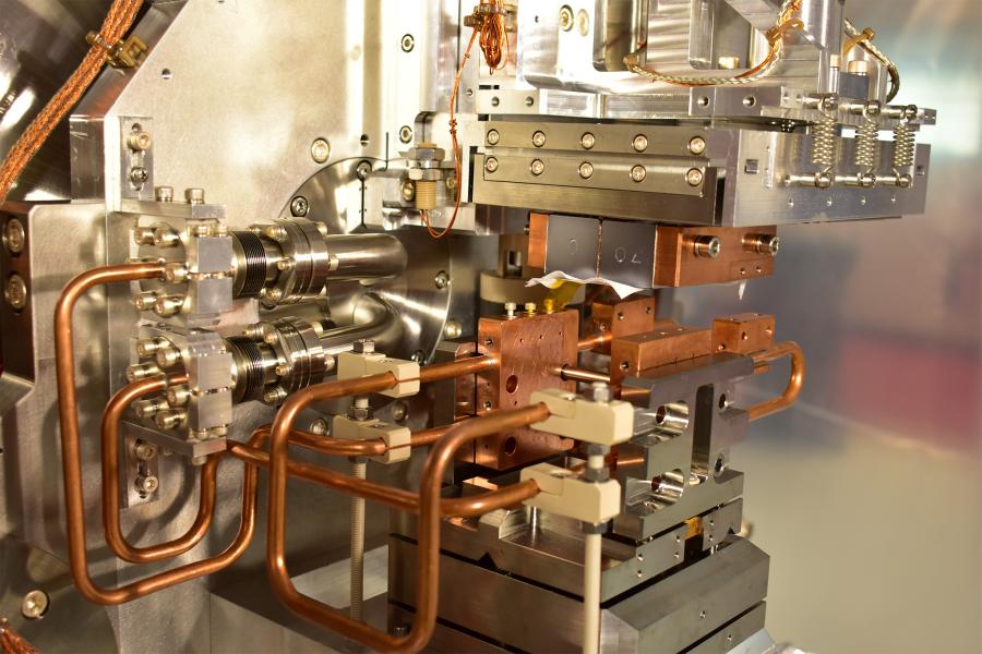
MEX1 DCM configuration showing the second crystal set installed. The lower surface of the crystals is covered with a wipe for protection.
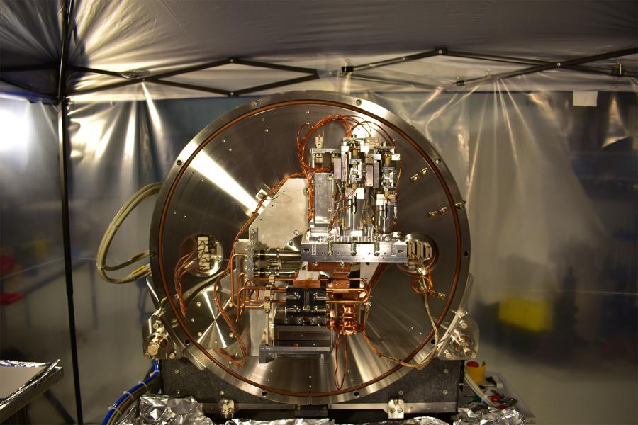
MEX1 with both crystal sets installed. The crystals are just below the centre of the image, and are covered with white lab wipes.
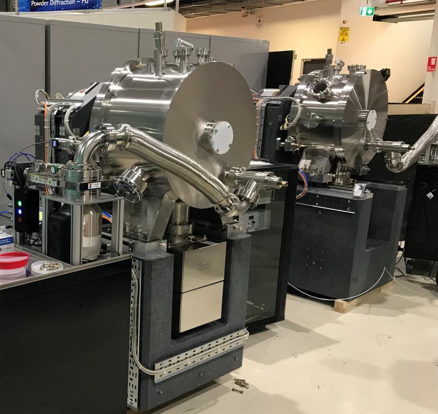
The final product: MEX1 and MEX2 DCMs completed and with their covers on.
In other June news, building of the MEX user cabin is now complete. The user cabin sits directly next to the hutches and is where users and Beamline Scientists will be located while operating the beamline. Check out the last stages of construction in the video below.
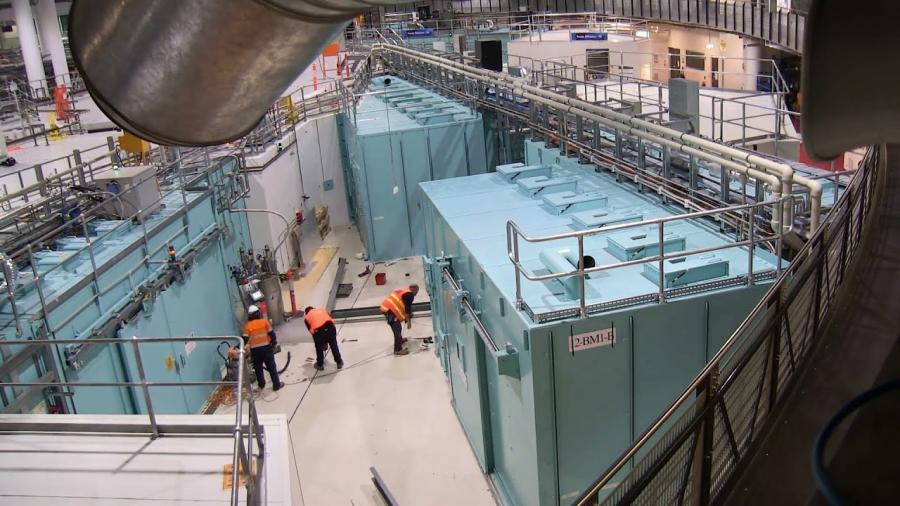
Video showing the construction of the MEX user cabins.
May 2021
May has been a busy month here at MEX. The final components of our hutches arrived and construction on them has now been completed. See the video below for the construction process following on from the March update.
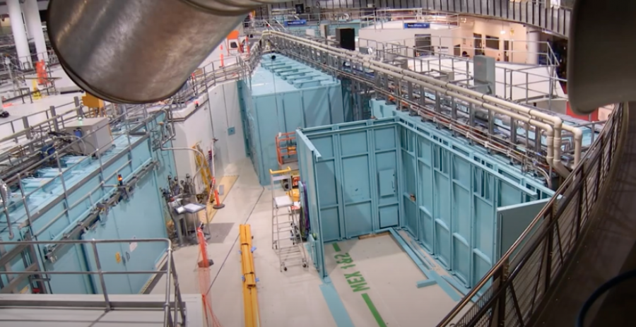
These hutches are now ready for utilities to be installed. Construction works will now focus on the user cabins, which our future users will soon(ish) become very familiar with as this is where they'll work when they come to use the Beamlines.
Our Controls Engineers have had a fruitful month working on our crystal spectrometer (check out the March update for more info). They now have all all the motors for the spectrometer working together. You can be mesmerised by the fruits of their labour in the image below.
Image showing each of the stages of the MEX crystal spectrometer moving together.
Now you'd better go stock up on popcorn because with the first components of the Beamline being assembled in the coming weeks, the June update is going to be a big one!
April 2021
This month we celebrated the arrival of our newest MEX Beamline Scientist, Dr Krystina Lamb. With Krystina's arrival, we now have a full MEX team!
We thought this would be a good time to introduce you to the gang working to get MEX operational for our future user community.
The MEX build is managed by our Project Manager and fearless leader, Mr Mohamed Elrabiey. Mohamed ensures the project is progressing on time and on budget, and coordinates parts and people coming in from all over the world (in COVID times!).
MEX's Mechanical Engineer, Mr Ben Pocock, leads the designing and sourcing of Beamline parts. He is responsible for all the mechanical components of the Beamline.
Mr Ben Baldwinson is our Senior Controls Engineer, and works with fellow Controls Engineers Mr Danny Wong and Mr Ross Hogan to ensure the Beamline components operate as they should. The Controls engineers bridge the gap between the Beamline hardware and software.
Mr Dion Curic is MEX's Mechanical Technician. He makes, designs, and assembles Beamline components.
Our Scientific Computing team, Senior Scientific Software Engineer Dr Letizia Sammut and Scientific Software Engineer Mr Alex Palma, design and implement all the user-focused Beamline software components. Mr Andrew Starritt is constructing the Graphical User Interfaces for Synchrotron scientists to commission and operate the Beamline.
Finally, meet the MEX Beamline Science team! Once the Beamline is operating, the Science team will be running the Beamline, training and assisting users, and troubleshooting any Beamline issues. For now, the scientists are ordering Beamline components and directing Beamline specifications. This team is led by Dr Chris Glover, and consists of Beamline Scientists Dr Jeremy Wykes, Dr Simon James, Dr Emily Finch, and Dr Krystina Lamb. We look forward to meeting you when MEX opens to users in 2022!
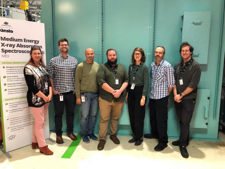
Photograph of some of the MEX team in front of the newly constructed MEX hutches. Left to right: Krystina Lamb, Simon James, Chris Glover, Ben Pocock, Emily Finch, Jeremy Wykes, Ross Hogan.
March 2021
Building has begun!
Our trusty builders have commenced assembly of the MEX Beamline hutches. The hutches walls are blue to differentiate from the white user cabins, and contain lead to shield radiation.
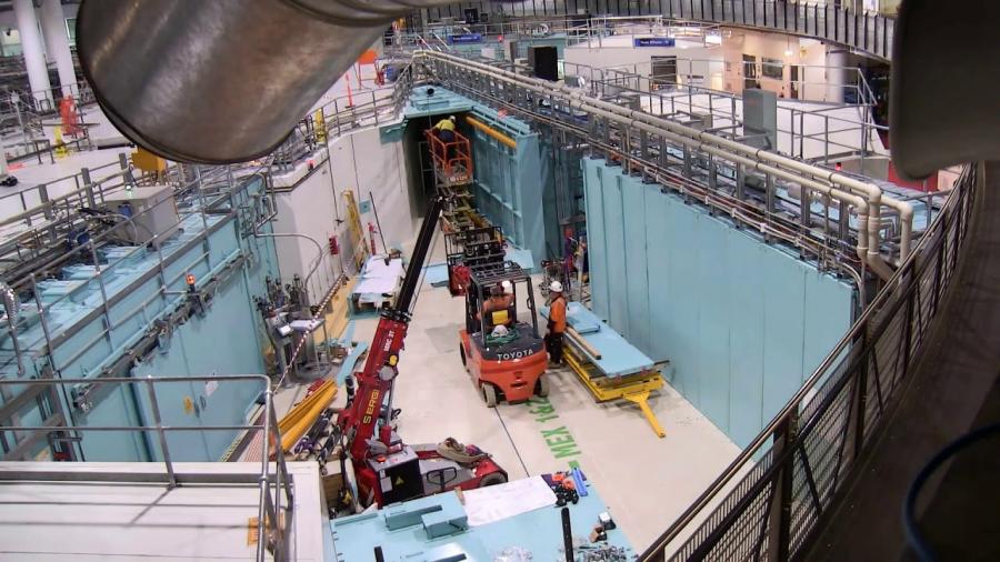
Once the hutches are built, the mechanical and electrical components will be installed.
Behind the scenes, parts are trickling in from around the world ready for installation in the hutches. One of our most exciting recent arrivals is the crystal spectrometer, which will produce high energy resolution spectra to allow us to discern detail that would otherwise be obscured. This piece of kit is an exciting new technology for our Synchrotron users!
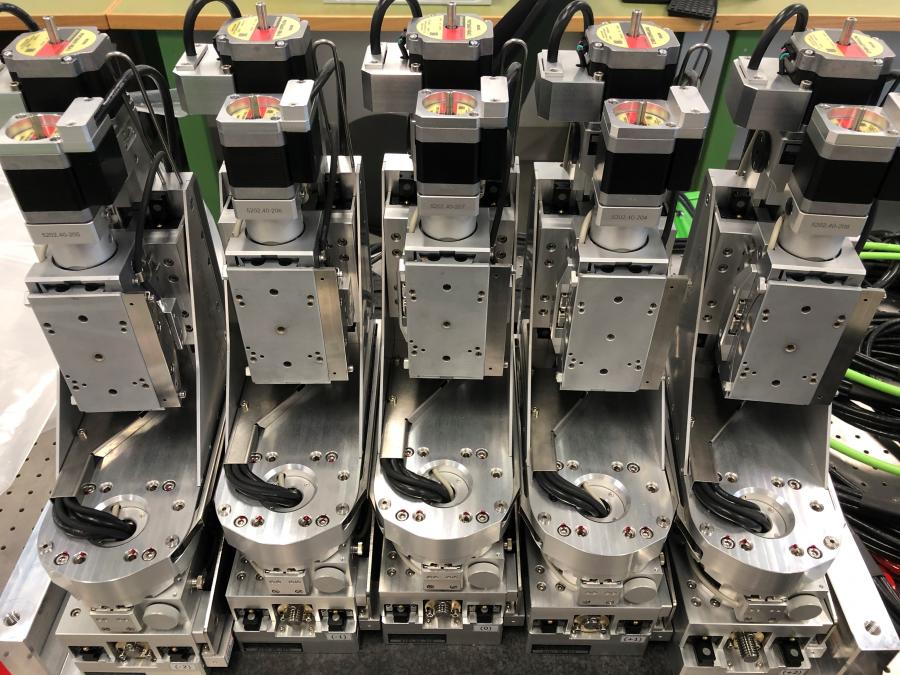
The main optical components for the Beamline are currently undergoing factory acceptance testing before being shipped in the coming days and weeks.
Stay tuned for monthly updates as parts continue to arrive and activity ramps up!
You can now apply for beam time on MEX1 and MEX2!
MEX1 and MEX2 are new beamlines which came online in late 2022 and early 2023. We will be progressively rolling out their capabilities over the coming months. For more information, including a schedule of capabilities, see the Australian Synchrotron User Wiki.
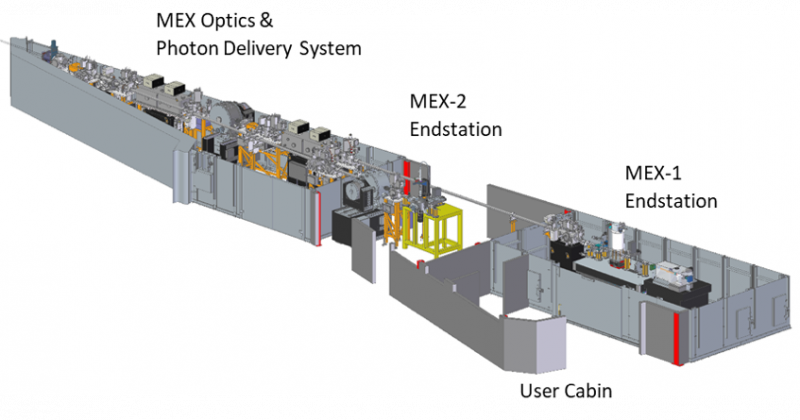
The MEX-1 and MEX-2 beamline utilises one bend magnet as the source and will operate independently and has been designed to be experimentally flexible, extending and complementing the suite of the X-ray spectroscopy techniques already available at the Australian Synchrotron.
At MEX, the instrumental capabilities are focused on:
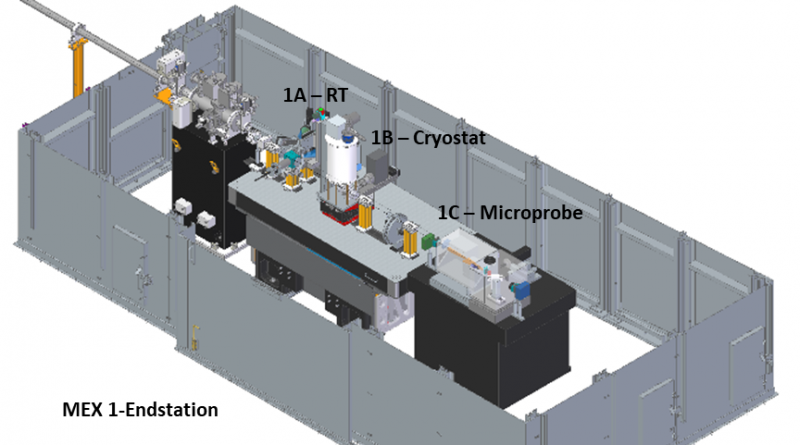
This beamline will offer monochromatic X-rays (from a Si<111> double crystal monochromator) at one of three endstations, with variable beam sizes at a fixed height from the floor. The different MEX-1 endstations will not be able to operate simultaneously.
Multiple modes of operation:
This branch line will offer low energy monochromatic X-rays – from either InSb<111> or Si<111> – double crystal monochromators at a fixed position and with a maximum beam size of 2 x 5 mm2. Slits can be used to reduce the beam profile on the sample. Sample environment: variable temperature (~10 – 500 K), helium gas atmosphere and/or vacuum.
Multiple modes of operation:
The MEX beamlines have been designed to offer a range of capabilities for undertaking XAS measurements across a wide range of scientific applications, with a particular emphasis on in situ and in operando experiments. Collecting high-quality data while minimising the dose delivered to the sample is expected to be another strength. MEX will also bring the new and powerful tool for chemical research, HERFD-XAS, to the Australian Synchrotron, opening new avenues for material characterisation.
In soils and minerals, silicates, phosphates, sulfur and chloride ions are common, and X-ray absorption spectroscopy of these ions can reveal the chemical environment in which they are present. When combined with the XAS on metals present in the same milieu, superior definition of the chemical speciation and spatial distributions will be possible compared to relying on a metal K-edge XAS alone. Such information is invaluable for the study of soil salination, the efficacy of fertilisers in agriculture, plant growth and micro-nutrient transport, as well as the distribution and bioavailability of essential micro-nutrients in food.
The target energy range of the MEX beamlines will open up numerous avenues of research into advanced materials.
Studies of numerous pollutants in the environment and their decomposition are readily studies using the MEX beamlines. Examples include:
By making use of non-ambient sample environments, experiments designed to reveal the speciation of complexes under hydrothermal conditions or lead to a better understanding of geochemical process like the transport and deposition of precious metals through the earth’s crust can be undertaken. Examples include:
Many of the pigments used in artworks and prevalent in archaeological artefacts contain metals as well as sulfur-containing, chloride and phosphate ions.
When MEX studies are combined with the dating of samples, information such as ancient cultural practices and trade associations lead to important understandings of many aspects of the development of civilisation and the preservation of cultural heritage worldwide
Number of Beamlines | MEX-1: monochromatic, 2.1 – 13.6 keV MEX-2: monochromatic 1.7 – 3.5 keV | |
Number of Endstations | MEX-1: 3 MEX-2: 1 | |
Source |
| Bending magnet (BM12-1) |
|
|
|
Optics | MEX-1 |
|
| Mirror 1 (M1) – Vertical collimation
| - Fixed, flat Si substrate - Cylindrically bent - Bounce up (4.75 mrad) - Exchangeable C, Ni and Rh stripes |
| Double crystal monochromators (DCM) | - Si<111>, ?E/E ≈ 1.4 ×10⁻⁴ - Crystal azimuth (?) 0° and 30° - Vertical bounce |
| Mirror 2 (M2) – Vertical and horizontal focussing | - Cylindrical + flat Si substrate - Elliptically bent - Bounce down (4.75 mrad) - Exchangeable C and Rh stripes |
|
|
|
| MEX-2 |
|
| Front end mirror (M1) – Vertical collimation | - Fixed sagittal cylinder - Horizontal bounce (15 mrad) - Cr stripe |
| Mirror 2 (M2) – Horizontal collimating | - Fixed cylinder - Horizontal bounce (15 mrad) - Cr stripe |
| Double crystal monochromators (DCM) | - Si<111>, ?E/E ≈ 1.4 ×10⁻⁴ - InSb<111>, ?E/E ≈ 3 ×10⁻⁴ - Crystal azimuth (?) 0° - Vertical bounce |
|
|
|
Endstations | MEX -1 |
|
| XAFS Station | - EXAFS/XANES - 3.5 – 13.6 keV - Transmission and florescence (4 el. Si SDD) - Sample temperature 10 – 300 K (in He(g)) - HERFD-XAS: > 0.5 m Johann-type crystal analyser > 5x spherically bent crystals. See here for further information on crystal cuts and accessible emission lines
- Non-ambient sample environments - Beam size 0.15 – 0.5 x 1.8 – 7 mm2 - Flux 3 x 10¹¹ ph-1 sec-1 |
| Microprobe station | - µEXAFS/µXANES - 2.1 – 13.6 keV - 3 el. Si SDD - 2-moment KB mirror system - Exchangeable C and Rh stripes - Beam size: 2 – >10 µm - Flux 108 ph-1 sec-1 µm2 - Sample in He(g) - Sample temperature 80 – 500 K - Scan range < 80 × 80 mm |
| MEX-2 (“Low energy station”) | EXAFS/XANES - 1.7 – 3.5 keV - Transmission, drain current and florescence (4 el. Si SDD) - HERFD-XAS: >Dispersive refocusing Rowland geometry >Single cylindrically bent, Si<111> Johann analyser - Beam size 0.1 – 2 x 0.1 – 5 mm2 - Flux: Si<111> 5 x 10¹⁰ ph-1 sec-1 InSb<111> 1 x 10¹¹ ph-1 sec-1 - Sample in He(g) or vacuum - Sample temperature 10 – 300 K |
Date | Status |
2017 July | Project started |
2018 January | Investment Case (IC) approved and endorsed by ANSTO |
2018 June | Conceptual Design Report |
2019 March | PDS Contract Placed (Axilon, Germany) |
2019 July | Technical Design Report |
2019 August | DCM (X2) Contract Placed (IDT, UK). |
2020 February | Crystal Spectrometer Order Placed (Huber, Germany) |
2021 Q1 - Q2 | Arrival of Crystal Spectrometer and DCMs on site |
2021 Q1 - Q2 | Installation of Hutches and User cabin |
2021 Q2 - Q3 | Installation of PDS and DCMs |
2021 Q3 - Q4 | Installation of endstations |
2022 Q1 | Hot commissioning begins, includes expert users |
2022 Q3 | First User Experiments; Beamline fully commissioned over the next 12 months |
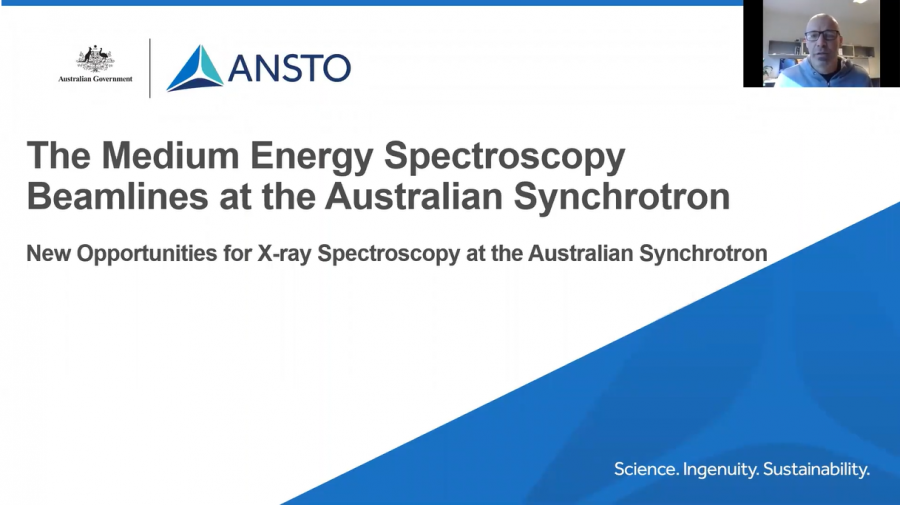
The talk below from MEX Lead Scientist Dr Chris Glover provides a summary of the MEX Beamline equipment and capabilities. Chris recorded this talk for the XAFS 2021 online conference.
 |  |  |