Infrared microspectroscopy
Introduction
The combination of high brilliance and collimation of the synchrotron beam through a Bruker VERTEX 80v Fourier transform infrared (FTIR) spectrometer and into the Hyperion 3000 IR microscope enables high signal-to-noise ratios at diffraction limited spatial resolutions between 3-8 μm, making the Infrared Microspectroscopy (IRM) beamline ideally suited to the analysis of microscopic samples e.g. small particles, thin films and layers within complex matrices, as well as single cells and complex biological systems.
Infrared microspectroscopy is a non-destructive method used to gain chemical and structural information for a diverse range of samples from biological, biomedical, materials, forensic and food sciences as well as cultural heritage and geology to name a few. The benefit of FTIR microspectroscopy compared to traditional FTIR spectroscopy, is that spatially resolved chemical information is acquired, enabling the production of "heat" maps that visually demonstrate the spatial distribution of specific chemical functionalities. The benefits of the synchrotron radiation sourced infrared light compared to traditional globar source is the smaller focused beam spot enhances the spatial resolution achievable and signal to noise of the resulting spectra.
In addition to the online Hyperion 3000 microscope, an offline Hyperion 3000 microscope attached to a Bruker VERTEX V70 spectrometer (using traditional globar source IR), is also available. Both microscopes are equipped with a single point MCT detector and a 64 × 64 focal plane array detector (FPA), enabling the rapid acquisition of high resolution chemical maps. Users can request access to the offline microscope during their beamtime, if required.
Both the online and offline microscopes can be operated in the following modes of data collection and can be coupled to a number of different accessories to assist with the wide variety of experiments conducted at the beamline:
- Transmission
- macro Attenuated Total Reflectance (ATR)
- Reflectance
- Grazing Incidence
In each of the above modes a motorised stage allows the point-by-point collection (raster mapping) of individual spectra from a sample, to create the spatially resolved “spectral maps”.
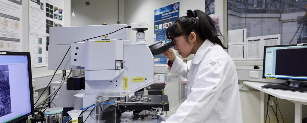
Top: An external view of the IR Microspectroscopy cabin. Bottom left: An internal view of the cabin with spectrometer and microscope. Bottom right: The Hyperion 3000 microscope
Samples analysed on the IRM beamline
The IRM beamline analyses samples from across a broad range of research and industry applications, including:
Biological and biomedical science
Synchrotron radiation infrared microspectroscopy is particularly useful for monitoring molecular information of single cells and distinguishing complex biological tissue sections at a sub-cellular resolution. Many researchers are seeking single cell information, whether to reveal biological variability, monitor differences in cell cycles, stem cell differentiation or the cellular uptake and resulting biochemical and structural effects of various chemicals and therapeutics to highlight a few examples, where traditional Globar sourced IR techniques would otherwise provide averaged information from a cell population.
For biological tissue, embedding and microtomed sections placed on an IR transmissive window such as CaF2 are commonly used for sample preparation. Alternatively, the embedded biological material may be microtomed and analysed using ATR-FTIR. Traditional sample preparation for cells involves fixing pre-treated cells onto an IR transmissive window for analysis in transmission mode. More recently, it is possible to study live cells making use of the flow cells available.
Types of samples include: salvia and dental samples, bone, single cells, tissue sections, pharmaceuticals
Materials and interfacial science
The information gained from infrared microspectroscopy, such as the heterogeneity of surface and bulk chemistries, orientation and structural conformation, and stability under conditions of interest, make its utility in materials science invaluable. Furthermore, temporally resolved rapid scan IR is now available, which will have particular benefits for material scientists who want to probe the kinetic chemical and structural effects to the sample as a result to exposure of a particular stimuli.
Methods for preparation and analysis of materials varies greatly depending on material properties and mode of analysis.
Types of samples include: natural and synthetic fibres (carbon fiber, silk, spider web, wool), polymeric material, nanoparticle coatings, membranes
Food science
The use of FTIR microspectroscopy in food sciences enables one to probe various chemical and structural information relating to parameters such as food processing conditions, chemical composition, influence of storage and shelf life, probe for adulteration and aid in the development and optimisation of potentially new products. We have temperature controlled stage accessories that enable measurements to be performed at refrigerated storage conditions, and use of the macro-ATR enables analysis without any sample preparation.
Examples of food types include: cheeses, yoghurt, bread, chia seed oil microcapsules
Geological and environmental sciences
The infrared microscopy beamline is particularly suited for the analysis of geological and environment samples. Along with 'dry' samples used in transmission and macro-ATR studies, the option to work in an aqueous medium using one of the beamline's liquid cell sample chambers is also available. The ability to work in liquid is important for investigating aquatic organisms and how they interact with their surroundings (e.g. chemical changes in the cell in response to a changing aqueous environment). A diverse collection of samples have been studied at the beamline, including algae (liquid cell), fungal infestation of leaves, occlusions in rock samples, and the symbiont relationship of corals.
Examples of samples include: soil samples, rocks, volcanic ash, plants, algae, coral, and pollutants.
Zoology and entomology
Analysis of insect wings is well established on the infrared microspectroscopy beamline, along with reptile skin. A variety of information can be obtained such as chemical and spatial composition and spatial distribution, low fouling and antibacterial properties, influence of geographical distribution, and effects of environmental pollutants. Use of the macro-ATR attachment is popular for insect wing analysis due to the ability to analyse these without any sample preparation or embedding required.
Forensic science
The infrared microspectroscopy beamline can been used for research in the forensic science arena for such purposes as fingerprint analysis, drug detection, human hair fiber analysis, and analysis of the authenticity of multilayered polymeric security cards to name a few examples. The technique is of particular use to forensic science where spatially correlating specific chemical information using non-destructive methods is of critical importance.
History and cultural heritage
The IRM beamline has been used by those involved in cultural heritage to probe a range of factors including the chemical analysis of pigments, paints, material etc in order to determine the historical period and geographical location that a historical object might originate from, as well as product authenticity or adulteration.
Selected publications from the beamline refined by research focus can be found on our technical website:
IRM publications by research focus
A full list of publications from the beamline can be found on the Portal:

